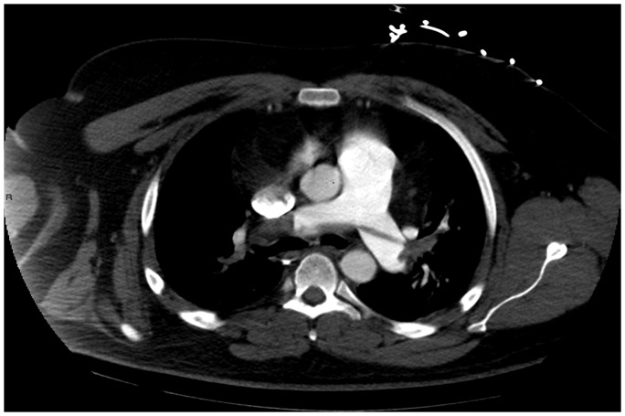
Figure 1.
Contrast-enhanced CT pulmonary angiogram at the level of the pulmonary trunk bifurcation demonstrates multiple low attenuation filling defects at the bifurcation and within the right and left pulmonary arteries, left interlobar and superior lingular artery in keeping with saddle embolus and multiple bilateral pulmonary emboli. The pulmonary trunk is dilated (36mm) which is concordant with concomitant pulmonary hypertension. Images not included demonstrate emboli involving all segments of both lungs and CT findings of right heart strain.

