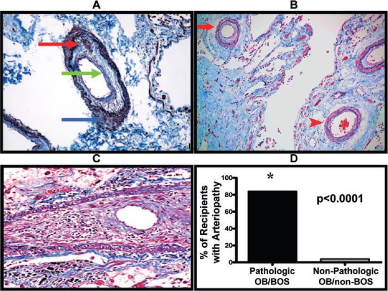
Figure 2. Pulmonary allograft arteriopathy occurs during pathologic OB/BOS.

(A) Representative example of a pulmonary artery demonstrating arteriopathy with moderate intimal fibrosis and mononuclear cell infiltration (red and green arrows). Also note the media with mild matrix deposition and no smooth muscle hypertrophy (blue arrow) (Masson’s trichrome/elastic Verhoeff van Gieson stain, original magnification 100×). (B) Representative example of the heterogeneous distribution of the arteriopathy as highlighted by concentric arteriopathy (red arrow) in the same field as a relatively normal artery (red arrowhead); (Masson’s trichrome/elastic Verhoeff van Gieson stain, original magnification 100×). (C) Representative example of a pulmonary artery with severe stenosis due to massive intimal fibrosis and mononuclear cell infiltration. Note the striking medial smooth muscle atrophy; (Masson’s trichrome/elastic Verhoeff van Gieson, original magnification 200×). (D) Lung transplant recipients with pathologic OB/BOS have significantly more arteriopathy as compared to lung transplant recipients without pathologic OB/BOS.
