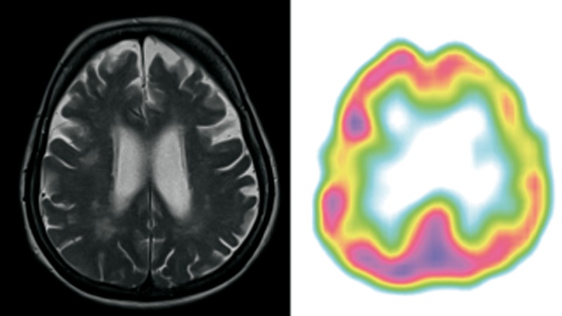
Figure 3.

Patient 3 – clinical diagnosis – lvPPA, MRI – lvPPA/nfvPPA bilateral atrophy of frontal lobes and subtle atrophy of parietal lobe. The ventricles were enlarged in proportion to atrophy with left-sided predominance. SPECT – lvPPA predominant left posterior frontal, perisylvian and parietal hypoperfusion.
