Figure 3.
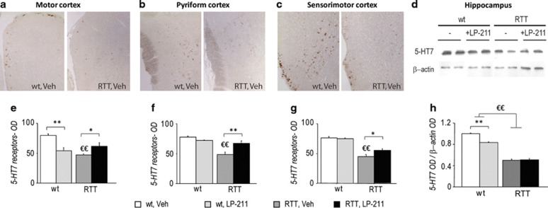
5-HT7 receptor density is reduced in the brain of RTT mice. (a–c) Representative photomicrographs showing the increase in immunoreactivity in the Motor (a), Pyriform (b), and Sensorimotor (c) cortical brain areas of wild-type (left panels) compared with RTT mice (right panels). All images are magnified equally ( × 20). (d) Examples of western blot analysis (representing a summarized view corresponding to two animals per group) of 5-HT7 and β-actin proteins in hippocampi of RTT and wt mice in control conditions (Veh) or treated with LP-211. (e–g) Histograms illustrate the semiquantitative evaluation of 5 HT7 receptor immunoreactivity in cortical brain areas. Values are expressed as means of optical density (OD) values of the immunoperoxidase labeling. (h) Histogram illustrates the semiquantitative densitometric analysis of western blot analysis in the hippocampus, obtained by OD of protein bands normalized with OD of β-actin bands. OD ratios are expressed as the average fold increase vs wt controls. LP-211 or vehicle were i.p.-administered for 7 consecutive days. Data are mean±SEM. Statistical significance was calculated by two-way ANOVA with Tukey's post hoc test. €, wt; Veh vs RTT, Veh; p<0.05; *, RTT, Veh vs RTT, LP-211, p<0.05, after post hoc comparisons on the genotype*treatment interaction.

