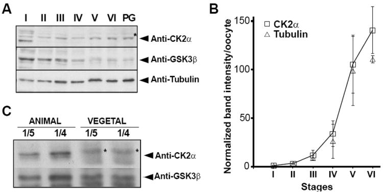
Figure 3. CK2α protein is enriched in the animal hemisphere of stage VI Xenopus oocytes.

(A) Expression of CK2 proteins during Xenopus oogenesis. Extracts were prepared from oocytes stages I, II, III, IV, V, VI, and stage VI treated with progesterone (PG). The volume equivalent of 0.2 stage VI oocytes was subjected to immunoblotting. Thus, we loaded, 22 stage I oocytes, 6.8 stage II oocytes, 2.5 stage III oocytes, 0.7 stage IV oocytes, 0.3 stage V oocytes and 0.2 stage VI oocytes. All immunoblots were repeated three times. The asterisk (*) indicates previously identified non-specific cross-reactive proteins in Xenopus extracts [15].
(B) Graph representing the data (band intensity) from the three independent experiments as in Figure 3A. Protein band intensity was divided by the number of oocytes loaded to obtain the band intensitity per oocyte, normalized to stage I oocyte (stage I =1), and represented as mean ±S.D.
(C) Immunoblot analysis of CK2 protein levels in 10 pooled stage VI animal or vegetal halves. One fifth (1/5, 0.2 μl) and one fourth (1/4, 0.25 μl) of oocyte lysate were loaded for immunoblot analysis. The asterisk (*) indicates a non-specific cross-reactive protein in Xenopus extracts [15]. CK2α protein is animally enriched at stage VI of oogenesis, while other tested proteins are not. This is a representative experiment out of three with identical results.
