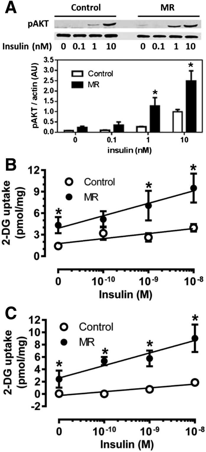
Figure 3.
Effect of dietary MR on ex vivo insulin signaling and insulin-stimulated glucose uptake in isolated white and brown adipocytes. Epididymal white adipocytes or brown adipocytes were isolated after 8 weeks of dietary MR and incubated with increments of insulin for 5 min (Akt) or 15 min ([3H]-2-DG) before measurement of Akt phosphorylation (A) and [3H]-2-DG uptake (B) in white adipocytes or [3H]-2-DG uptake in brown adipocytes (C) as described in Research Design and Methods. Scanning densitometry was used to quantitate expression levels for each protein. Means ± SEM are representative of three independent experiments. *Mean responses at each insulin concentration differ from their corresponding controls at P < 0.05.

