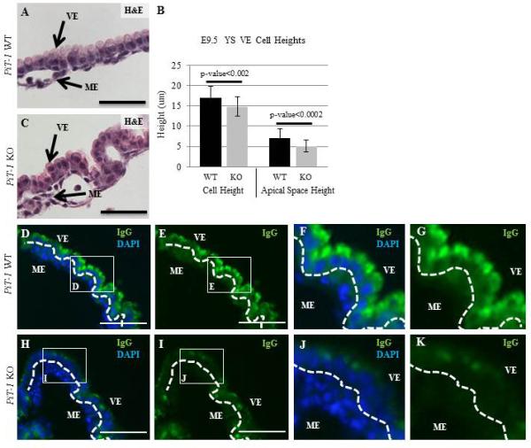
Figure 5. Abnormal Morphology and IgG Localization in PiT-1 KO YS VE.

Quantification of cell size and nuclear localization relative to the apical membrane reveals that PiT-1 KO VE cells are smaller due to a decrease in the apical space (A-C). There is a large reduction in IgG accumulation in the PiT-1 KO YS VE (H-K), compared to controls (D-G). Scale bars=100um.
