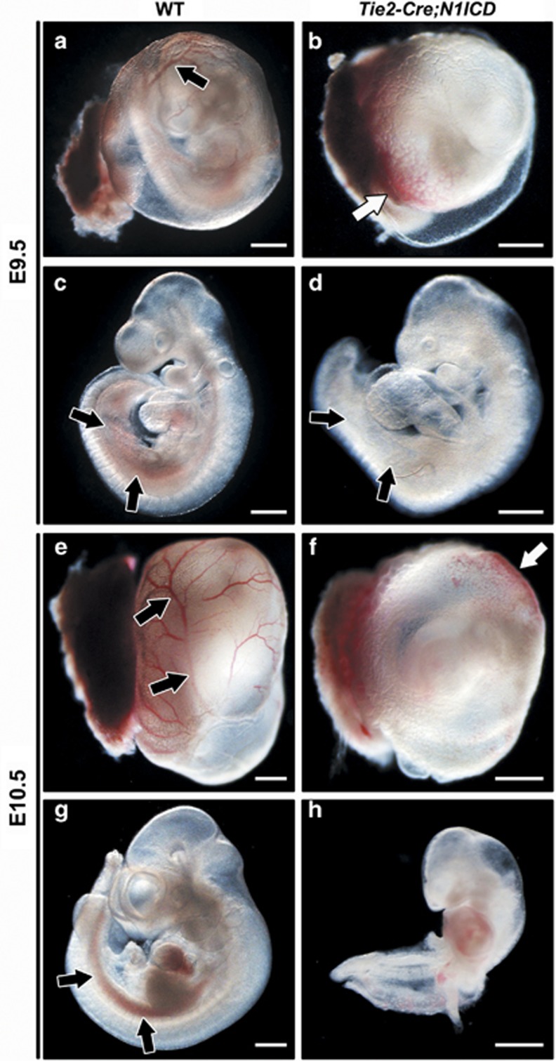
Figure 1.
Constitutive Notch1 activation on Tie2+ progenitor cells impairs hematopoietic development. WT and Tie2-Cre;N1ICD embryos (left and right panels, respectively) at E9.5 (a–d) and E10.5 (e–h), with the YS (a, b, e and f) and without it (c, d, g and h). (a, b) Morphology of the YS of WT and Tie2-Cre;N1ICD embryos. WT YS contains normally developed blood vessels (black arrow in a) that are absent in transgenic YS. Tie2-Cre;N1ICD YS is very pale, with hemoglobinized cells concentrated at one pole (white arrow in b). (c, d) Lateral views of WT and Tie2-Cre;N1ICD embryos. Black arrows indicate the P-Sp/AGM region with hemoglobinized content in WT, and the pale region in the transgenic embryo, indicative of defective definitive hematopoietic development. (e–h) At E10.5, defects in Tie2-Cre;N1ICD YS and embryos are more severe. Black arrows in (e) mark the well-developed WT YS blood vessels that are never seen in transgenic YS (f), which instead contains randomly located pockets of high hemoglobin content (white arrow). Lateral views of WT (g) and Tie2-Cre;N1ICD (h) embryos show the hemoglobinized dorsal aorta in the AGM region (black arrows) of a WT embryo that is absent in the transgenic embryo. Scale bars: 800 μm

