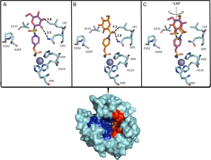
Figure 2.
(A) CA IX-mimic (cyan) and 5d. (A) Conformation 1 (purple) and (B) conformation 2 (orange) as they correlate to a surface representation depicting the location of 5d. Highlighted hydrophobic (red) and hydrophilic (blue) residues. Specific interactions and hydrogen bond distances (Å) are shown. (C) An overlay of each conformation of 5d. Note: there is a 170° rotation observed between sulfonamide bridges that distinguishes the two conformers. Figure was made using PyMol.16 Residues are as labeled (CA II numbering).

