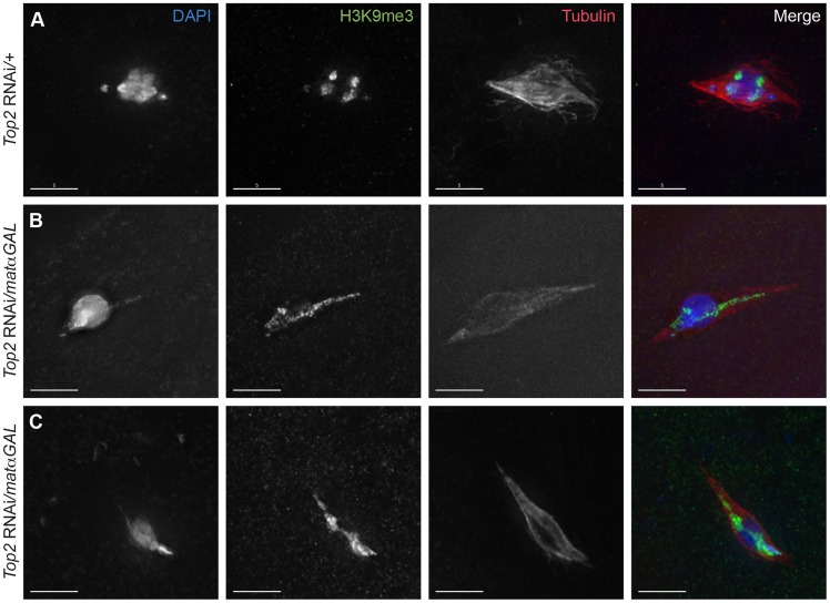
Figure 3. Aberrant DNA projections are composed of heterochromatin.
DNA is labeled with DAPI (blue), heterochromatin is labeled with an antibody recognizing histone 3 trimethylated on lysine 9 (H3K9me3) (green), and the spindle is labeled with an antibody recognizing α-tubulin (red). (A) Top2 RNAi/+ control oocyte with achiasmate 4th chromosomes that have moved towards the spindle poles. The 4th chromosomes and the centromeric regions of the chromosomes are labeled with H3K9me3 and are oriented towards opposite spindle poles. (B–C) Top2 RNAi/matαGAL oocytes containing abnormal DNA projections that are labeled with the H3K9me3 antibody. Additionally, the H3K9me3 antibody localization in the chromosome mass of both oocytes is not oriented towards opposite spindle poles. Images are projections of partial Z-stacks. Scale bars are 5 microns.

