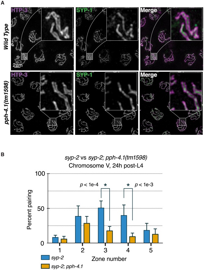
Figure 3. Canonical SC structure but reduced synapsis-independent pairing in pph-4.1 mutants.
(A) 3D-SIM images of pachytene nuclei immunostained for axial element HTP-3 (violet in merged image) and central element SYP-1 (green). Boxed insets at 5x higher magnification demonstrate position of SYP-1 between parallel tracks of HTP-3. (B) quantitation of SC-independent pairing of 5S rDNA loci in syp-2 and syp-2; pph-4.1 mutants. The percent of nuclei with paired foci in each of 5 zones (see Figure 2) is shown; error bars show SD. Six gonads were scored for each genotype. The total number of nuclei scored for zones 1–5 was as follows: syp-2 single mutant: 294, 322, 427, 417, 249; syp-2; pph-4.1 double mutant: 268, 281, 245, 295, 251.

