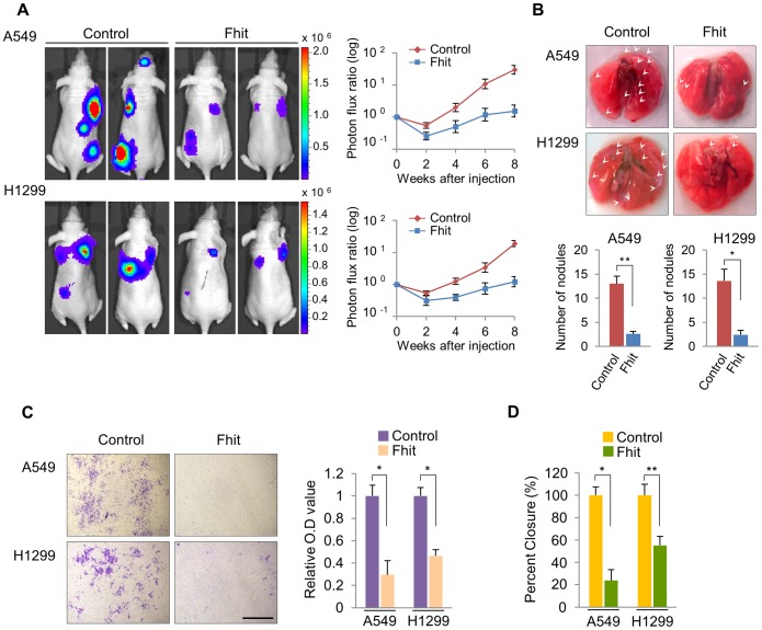
Figure 1. Elevated FHIT expression inhibits metastasis in human NSCLCs.
(A) Representative BLI plots of lung metastases in mice after injecting 1×10⁶ A549 or H1299 NSCLC cells stably expressing FHIT or a control vector; n = 4. (B) Lungs were extracted at 8 weeks after tail vein injection and the number of nodules was counted. (C) Representative images of FHIT-expressing or control cells that invaded through the filter and were stained with crystal violet. Scale bar, 40 µm. The results are means ± s.d, n = 4 experiments. *P<0.001. (D) Wound healing assay of confluent monolayer of FHIT-expressing or control A549 and H1299 cells at 0 h and 24 h. See Supplementary Fig. S3, that shows representative images of migration. *P<0.001, **P<0.0001.

