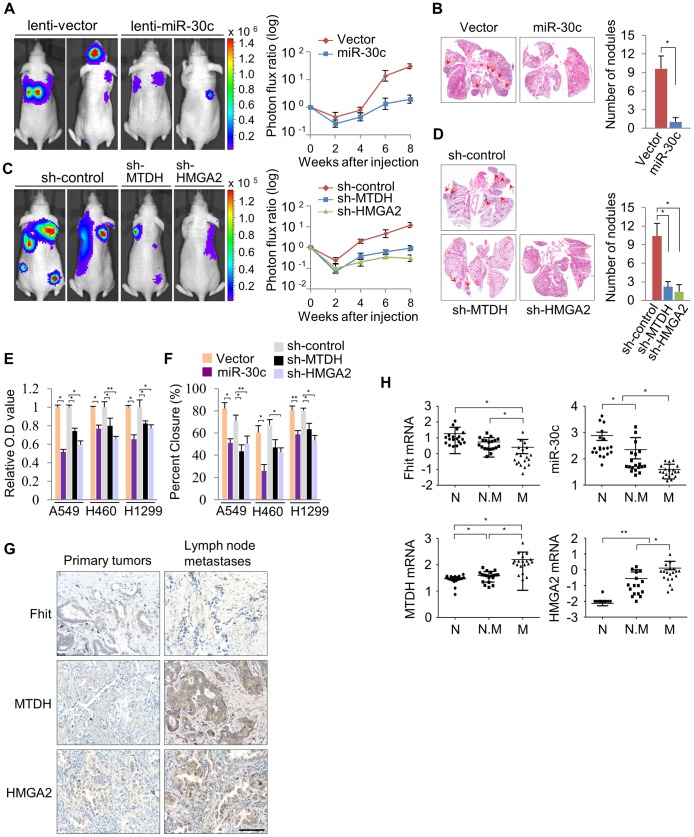
Figure 6. miR-30c inhibits metastasis through the suppression of MTDH and HMGA2 in NSCLCs.
(A) Representative BLI plots of lung metastasis of mice injected (IV) with 1×10⁶ A549 cells stably expressing miR-30c or a control vector; n = 4. (B) Representative H&E stained lung section. The arrows highlight metastatic nodules. *P<0.05 by Student's t-test. (C) Representative BLI plot of metastasis after IV injection with 1×10⁶ A549 cells after knockdown of MTDH, HMGA2, or control; n = 5. (D) Representative H&E stained lung section and the number of lung metastatic nodules. The arrows indicate metastatic nodules. *P<0.001. (E), (F) Invasion and migration assays in control and miR-30c-overexpressing cells, or control, MTDH and HMGA2 knockdown cells, respectively. *P<0.001 **P<0.01 by Student's t-test. (G) Immunohistochemistry assay for FHIT, MTDH and HMGA2 in primary lung tumors and their matched lymph node metastases. Scale bar, 100 µm. (H) The expression pattern of FHIT, miR-30c, MTDH, and HMGA2 in normal (N), non-metastatic (N.M) and metastatic tissues (M). *P<0.001 **P<0.05 by Student's t-test.

