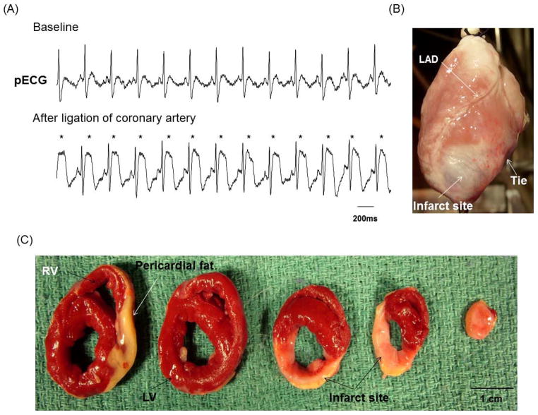
Figure 1.
Creation of chronic MI. A, After ligation of coronary artery, definite ST segment elevation (asterisks) was seen on the pseudo ECG (pECG). B, Photography of the anterior view of the infarcted heart showed white fibrotic area at apex of left ventricle (LV). C, The triphenyl tetrazolium chloride staining of the infarcted heart showed brisk red for surviving myocardium and white for infarcted myocardium. LAD, left anterior descending coronary artery; RV, right ventricle.

