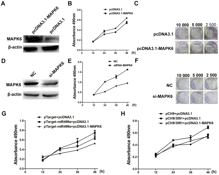
Figure 6. MiR499a promoted cell growth by reducing MAPK6.
(A) Western blot analysis showed the protein level of MAPK6 in SMMC-7721 cells transfected with overexpression plasmid pcDNA3.1-MAPK6 or control plasmid pcDNA3.1. (B) Cell proliferation was measured after transfected by pcDNA3.1 or pcDNA3.1-MAPK6 by MTS assay. (C) The colony formation assay showed the effects of MAPK6 overexpression on cell proliferation in concentration gradient. (D) The expression of MAPK6 in SMMC-7721 cells transfected with NC or si-MAPK6 was detected by Western blot. (E) Proliferation of cells transfected with si-MAPK6 was obviously increased as compared with NC transfection by MTS assay. (F) Representative pictures of colony formation assay of si-MAPK6 transfected SMMC-7721 cells. (G) The results of MTS assay illustrated the increase of cell proliferation transfected with pTarget-miR499a could be reduced by the overexpression of MAPK6. (H) The increase of cell proliferation by the overexpression of miR499a mediated by HBV could be inhibited though the overexpression of MAPK6. The results were measured by MTS assay. Statistically significant differences are indicated:*p<0.05, **p<0.01, Student's t test.

