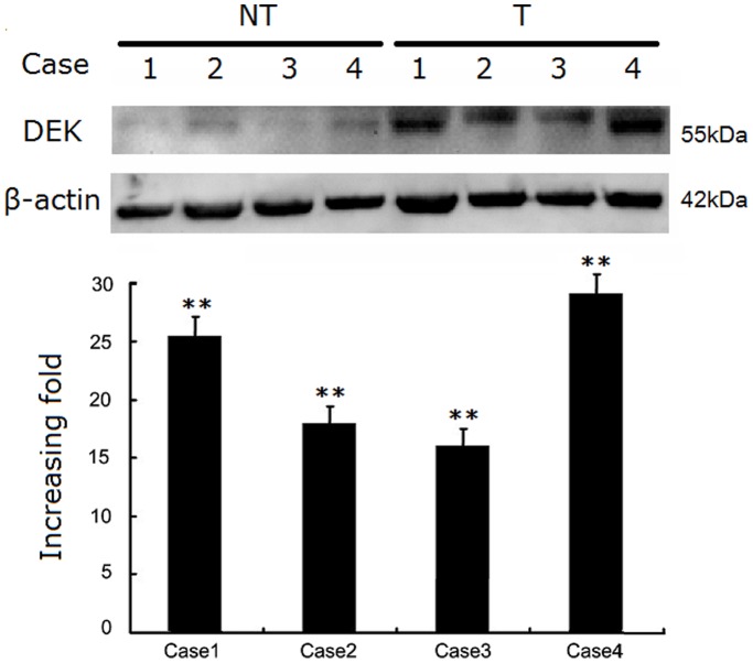
Figure 1. Western blotting analyses of the DEK protein.
(A) Images of western blots of DEK protein expression in four matched pairs of CRC (T) and adjacent non-tumor tissues (NT). (B) Relative T/NT ratios of DEK protein expression levels in paired CRC and adjacent non-tumor tissues are shown (increased fold change, **P<0.01).

