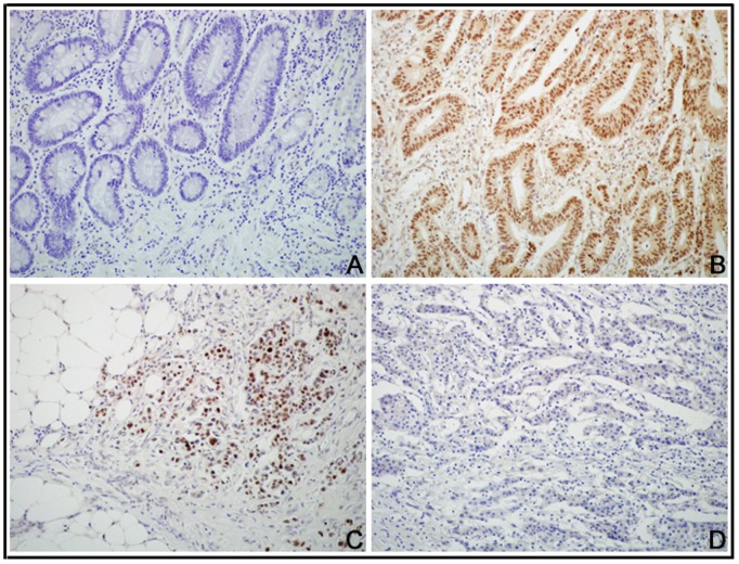
Figure 2. DEK protein expressed in CRC using IHC.
(A) DEK protein staining was negative in adjacent normal colon tissues. (B) DEK protein showed diffusely and strongly nuclear positive staining in late-stage CRC. (C) DEK protein is positive in invasive cancer cells in the serosa of the colon. (D) DEK protein is negative in CRC without metastasis.

