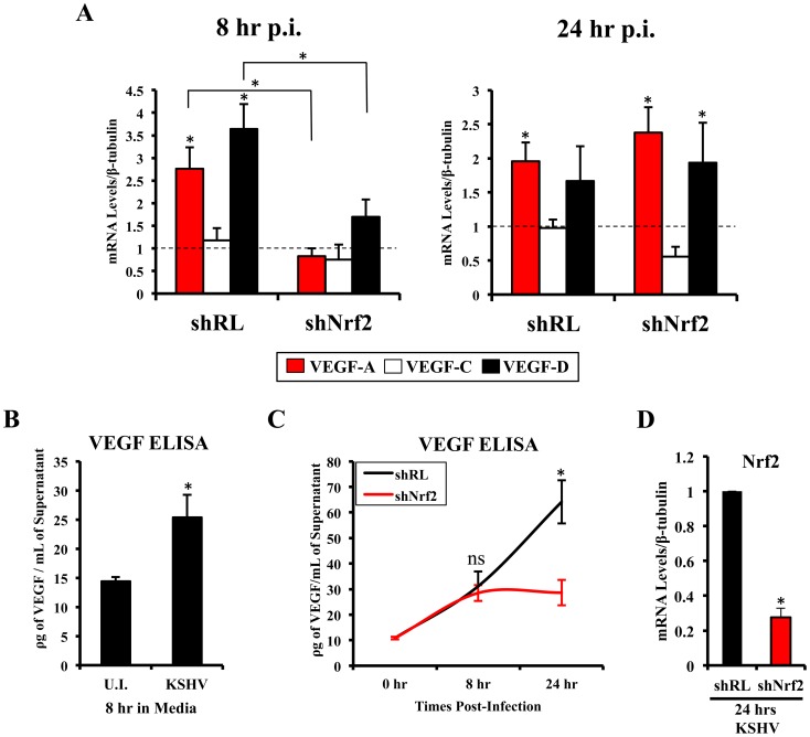
Figure 7. VEGF activity during KSHV infection and its dependence on Nrf2.
A) Starved HMVEC-d cells were infected with KSHV (20 DNA copies/cell) for 8 hr (left) and 24 hr (right) prior to real-time RT-PCR analysis for 3 members of the vascular endothelial growth factor (VEGF) family: VEGF-A (red), C (white) and D (black). β-tubulin was used as an endogenous house-keeping control and gene expression of uninfected cells (not shown) was arbitrarily set to 1 as depicted by the horizontal dashed line, while the bars indicate mean fold induction ± SD for 3 independent experiments. * = p<0.05 for each time/gene when compared to the respective U.I. control. B) VEGF165 ELISA (A-isoform) (Quantikine Kit – R&D) was performed on starved HMVEC-d cells infected with KSHV (20 DNA copies/cell) for 8 hr prior to supernatant collection and quantification of absolute levels using a standard curve. C) VEGF165 ELISA was performed in shRL and shNrf2 cells infected with KSHV (20 DNA copies/cell) for the indicated times post-infection. For panels B and C, values indicate the mean of supernatant protein concentrations ± SD for 3 independent replicates. D) Real-time RT-PCR of Nrf2 mRNA in conditions from panel C, to determine the efficiency of lentiviral knockdown of Nrf2. * = p<0.05.

