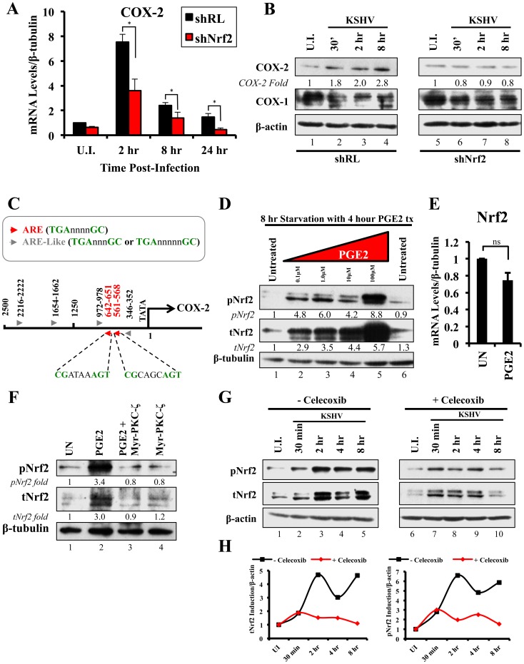
Figure 8. KSHV infection and the Nrf2-COX-2-PGE2 loop.
A) HMVEC-d cells were transduced with lentiviral vectors expressing shRL or shNrf2 for 24 hr, and then infected with KSHV (20 DNA copies/cell) for the indicated time points prior to real-time RT-PCR analysis for COX-2 (Cyclooxygenase-2). The U.I. shRL condition was arbitrarily set to 1 and the bars indicate fold induction ± SD for 5 independent experiments. β-tubulin was used as an endogenous control. * = p<0.05. B) shRL- and shNrf2-transduced HMVEC-d cells were infected with KSHV (20 DNA copies/cell) for the indicated time points and immunoblotted for COX-2 and COX-1. β-actin was used as a loading control and the fold induction relative to each U.I. condition (arbitrarily set to 1) is indicated. C) The human COX-2 promoter sequence (2500 base-pairs upstream of the transcriptional start site) was obtained from ensemble.org (Accession #: ENSG00000095303). Antioxidant Response Element (ARE) consensus sequences are indicated with red arrows, whereas ARE-like sequences are identified with grey arrows. D) Starved HMVEC-d cells were treated with an increasing concentration (0.1–100 µM) of Prostaglandin E2 (PGE2) for 4 hr prior to immunoblot analysis for pNrf2 and tNrf2. β-tubulin was used as loading control. E) Starved HMVEC-d cells were treated with 10 µM PGE2 for 4 hr prior to RNA extraction and real-time RT-PCR analysis of Nrf2 gene expression levels. β-tubulin was used as an endogenous control. The uninfected condition (U.I.) was arbitrarily set to 1, and the points indicate fold induction ± SD for 3 different replicates. ns = p>0.05. F) Starved HMVEC-d cells were pretreated with 10 µM Myristoylated-PKC-ζ pseudosubstrate for 1 hr before addition of PGE2 for an additional 4 hr and analysis by immunoblot assay for pNrf2 and tNrf2. β-tubulin was used as a loading control and the fold induction relative to each U.I. condition (arbitrarily set to 1) is indicated. G) HMVEC-d cells were pretreated with mock inhibitor or Celecoxib (10 µg/ml) for 2 hr prior to KSHV infection (20 DNA copies/cell) for the indicated time points and immunoblotted for pNrf2 and tNrf2. H) Graphical representation of the fold induction of pNrf2 and tNrf2 levels in Celecoxib- and mock (DMSO)-treated cells. Fold induction was normalized to β-actin and is relative to their respective U.I. condition, which was arbitrarily set to 1.

