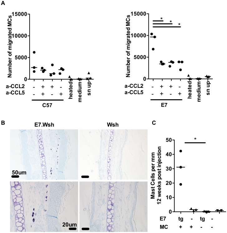
Figure 4. MCs migrate towards CCL2 and CCL5 and are recruited to E7 ear skin.
(A) 5 µm transwell assays were used to determine the mast cells chemotaxis towards chemokines. Graphs represent the number of migrated FcεRIα+ cKit+ BMCMCs towards C57 skin explant supernatants (left) or E7 explant supernatants (right) in the presence of anti-CCL2 and/or anti-CCL5 blocking antibodies used at 10 µg/ml. 3 independent experiments (B,C) Kit W-sh/W-sh (Wsh) (n = 3) and E7.Kit W-sh/W-sh (E7.Wsh) (n = 3) mice were reconstituted with 1.4×10⁷ BMCMCs i.v. or left untreated (E7.Wsh (n = 3) and Wsh (n = 2)). 12 weeks later, mice were culled and MCs were identified in ear skin by (B) toluidine blue staining (top, scale bar = 50 µm and bottom, scale bar = 20 µm) and expressed as (C) MC number per mm cartilage length. *p<0.01, by unpaired t-test for all indicated comparisons.

