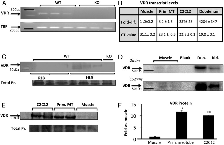
Figure 3.
Detection of VDR in skeletal muscle on PCR and Western blotting. A, On semiquantitative PCR, VDR transcript is detectable in quadriceps muscle from 3 adult WT mice. Muscle from 3 VDRKO mice was used as negative control. B, VDR transcript levels relative to whole muscle (ie, fold difference; VDR mRNA levels divided by that in whole muscle) and absolute CT values are listed (mean ± SEM, n = 3 per group). VDR transcript levels are markedly higher in duodenum (Duo.), the classic target of vitamin D action, compared with whole muscle and appreciably higher in muscle cell models compared with whole muscle. C, Using VDR-D6 antibody on Western blotting, VDR was detected in quadriceps muscle processed in HLB but hardly detected in samples processed in RLB (n = 9 per group, 60-μg protein/well, 7.2-fold higher detection; P < .05). For negative control, VDRKO muscle sample processed in HLB was used (n = 3). D, Compared with Duo. and kidney (Kid.), VDR in muscle required longer exposure time (15 min) and more protein loading (50 vs 10 μg per lane). Western blotting (E) and ImageJ densitometric quantitation (F) show that VDR expression (normalized for total protein on Coomassie) is approximately 10-fold higher in primary myotubes (Prim. MT) and C2C12 myotubes compared with whole muscle (n = 2 per group, P < .005, 20-μg protein/well). Samples used were processed in HLB. TBP, TATA box binding protein.

