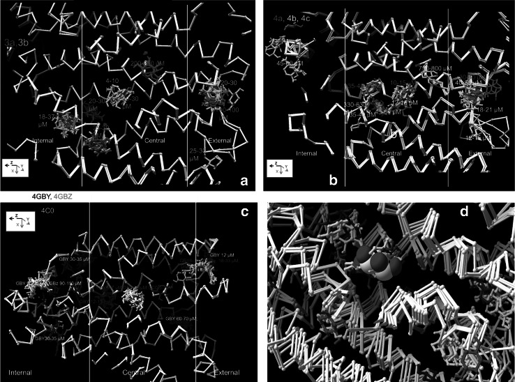
Fig. 2.
Side views showing xylose docking to a conformers 3a and 3b, b conformers 4a, 4b and 4c and c 4GBY, 4GBZ and 4GC0. The white vertical lines subdivide the transporter regions along the z axis approximately into external vestibular, central and internal vestibular zones; d The crystal docked xylose shown as the 3D rendered CPK coincides with the high affinity docking site as obtained with several XylE conformers shown as ball and stick forms (Color figure online)

