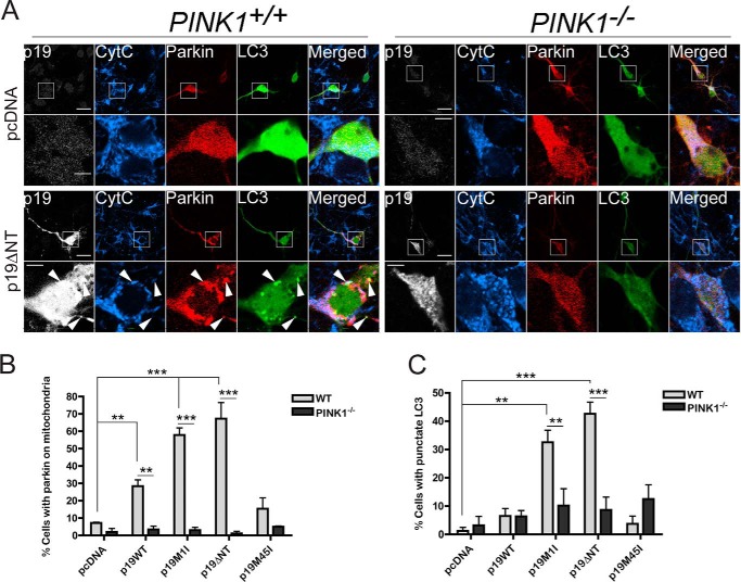
FIGURE 7.
smARF induces Parkin/PINK1-dependent mitophagy in neurons. A, cortical neurons generated from embryonic day 14 WT and PINK1−/− embryos were transfected with mCherry-Parkin, GFP-LC3, and either pcDNA, p19WT, p19M1I, p19M45I, or p19ΔNT constructs and processed 24 h later for immunofluorescence using cytochrome c (CytC) and p19 antibodies (only pcDNA and p19ΔNT shown). Scale bar, 20 μm for low power and 5 μm for zoom. Arrows, co-localization of p19ARF, mCherry-Parkin, cytochrome c, and GFP-LC3 (two top arrows) and co-localization of p19ARF, mCherry-Parkin, and GFP-LC3 without cytochrome c (bottom arrow). B, histogram showing the proportion of transfected cells with mCherry-Parkin co-localizing with the mitochondrial marker cytochrome c in WT versus PINK1−/− neurons. At least 25 neurons were counted per condition per experiment. All WT conditions are compared with PINK1−/− for each construct. All columns are also compared with pcDNA (separately for WT and PINK1−/− series) n = 3. **, p < 0.01; ***, p < 0.001. C, histogram showing the proportion of transfected cells with punctate GFP-LC3 staining in WT versus PINK1−/− neurons. At least 25 neurons were counted per condition per experiment. n = 3. All WT conditions are compared with PINK1−/− for each construct. All columns are also compared with pcDNA (separately for WT and PINK1−/− series). **, p < 0.01; ***, = p < 0.001. Error bars, S.E.

