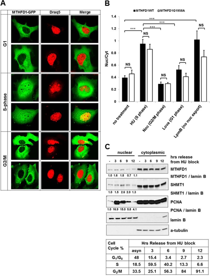
FIGURE 3.
MTHFD1 traffics to the nucleus in cell cycle-dependent manner. A, MTHFD1-GFP fusion protein localization within cell cycle-synchronized HeLa cells. HeLa cells were transfected with a plasmid encoding an MTHFD1-GFP fusion protein and arrested in G1, S, and G2/M phase. Two representative images for cells for the indicated phases of the cell cycle are shown. MTHFD1-GFP is colored green in the left panels with the nuclear stain Draq5 colored red on the middle panels; the merged images are in the right panels. B, quantitation of nuclear and cytoplasmic localization of MTHFD-GFP wt (black bars) and G1958A polymorphic variant (white bars) at each stage of the cell cycle, as described under “Experimental Procedures.” The ratios of fluorescence intensity in the nucleus to the cytosol were calculated for at least 20 individual cells per condition and are graphed as the mean ± S.E. The statistical significance p was assessed as a t test with Bonferroni correction for multiple comparisons and is represented as follows: NS (non-significant), ***, p < 0.001. Noc, nocodazole; Lpmb, leptomycin B. C, immunoblot analysis of MTHFD1 partitioning between the cytosolic and nuclear fractions in asynchronous HeLa cells, and in HeLa cells arrested in S phase and released into fresh medium for the indicated time points. S phase cell cycle arrest and release were confirmed by FACS analysis.

