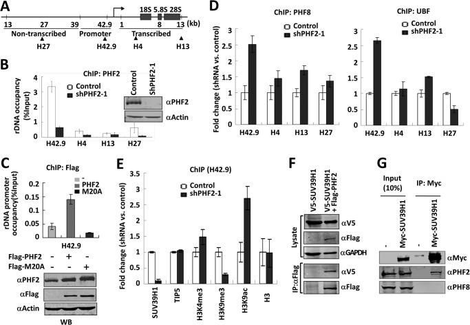
FIGURE 4.
PHF2 inhibits the binding of PHF8 and UBF and recruits SUV39H1 to the rDNA promoter. A, schematic diagram of a rDNA gene showing its promoter and non-transcribed and transcribed parts. Arrows point to different areas of rDNA paired by the four primer sets used in ChIP (see Table 1). B, ChIP analysis showing PHF2 is highly enriched in the rDNA gene promoter. ChIP analysis for PHF2 association at four different regions of the rDNA gene was performed with HeLa cells stably expressing either ShPHF2–1 or control shRNA. The results were presented as percentage of input. Error bars, S.D.; n = 3. The knockdown of PHF2 by shPHF2–1 was verified by Western blot analysis. C, PHF2 requires PHD domain to bind rDNA promoter. HeLa cells transfected with FLAG-PHF2 or FLAG-PHF2-M20A were subjected to a ChIP assay with anti-FLAG antibody, and the promoter association was analyzed. Error bars, S.D.; n = 3. A fraction of the cells were subjected to Western blot with PHF2, FLAG, and β-actin antibodies. D, knockdown of PHF2 led to increased association of PHF8 and UBF with the rDNA promoter. HeLa cells stably expressing either shPHF2–1 or control shRNA were subjected to ChIP analysis for PHF8 (left) or UBF (right). Note that the -fold changes in rDNA occupancy between ShPHF2-1-treated and control cells are shown. Error bars, S.D.; n = 3. E, knockdown of PHF2 led to decreased association of SUV39H1 and reduced H3K9me3 methylation at the rDNA promoter. A ChIP assay was performed as in C using antibodies as indicated. Error bars, S.D.; n = 3. F, co-immunoprecipitation of ectopically expressed SUV39H1 and PHF2. HeLa cells were transfected with V5-SUV39H1 alone or together with FLAG-PHF2, and immunoprecipitation was performed with FLAG antibody, followed by Western blot analysis with rabbit V5 and mouse FLAG and GAPDH antibodies. G, co-immunoprecipitation of ectopically expressed SUV39H1 with endogenous PHF2. HeLa cells were transfected with or without Myc-SUV39H1, and immunoprecipitations were performed with anti-Myc antibody, followed by Western blot analysis using anti-PHF2 or PHF8 antibody, as indicated.

