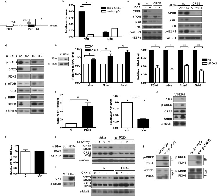
FIGURE 4.
PDK4 stimulates RHEB expression through stabilization of CREB. a, schematic illustration of the promoter regions of the mouse RHEB gene. E1 represents RHEB exon 1; the dark rectangle denotes a PBR, and the open rectangle indicates an NBR in the RHEB promoter. The arrow above the gene indicates the transcription start site. b, NIH/3T3 cells were subjected to ChIP assay using an anti-phospho-CREB antibody; normal rabbit IgG antibody served as the negative control. c, lysates of NIH/3T3 cells transfected with CREB siRNA or negative control siRNA treated with or without 20 mm DCA for 24 h and transfected with PDK4 or control plasmids were subjected to immunoblotting. d, lysates of NIH/3T3 cells transfected with two independent siRNAs for CREB or negative control siRNA were subjected to immunoblotting. e, total RNAs were extracted from NIH/3T3 cells transfected with PDK4 or control plasmids and NIH/3T3 cells transfected with siRNA for PDK4 or control siRNA for qRT-PCR. f, CREB antibody-immunoprecipitated DNA from PDK4-overexpressing and control NIH/3T3 cells and NIH/3T3 cells treated with 20 mm DCA for 24 h were PCR-amplified for regions indicated in a. g and h, total protein lysates and RNA were extracted from NIH/3T3 cells transfected with PDK4 or control plasmids for immunoblotting and qRT-PCR, respectively. i, immunoblotting of CREB in PDK4-overexpressing or control NIH/3T3 treated with MG-132 (10 μm). j, immunoblotting of CREB in NIH/3T3 with shPDK4 or shScr treated with CHX (25 μm). k, co-immunoprecipitation of endogenous proteins from NIH/3T3 cells and HEK293T cells. Immunoprecipitation (IP) was performed with anti-phospho-CREB or control IgG, and complexes were examined by immunoblotting with either phospho-CREB or PDK4 antibody. Co-immunoprecipitation and ChIP assay data were replicated in two independent experiments. Data represent mean ± S.E. of replicate real time PCR. nc indicates negative control. V indicates control plasmids. *, p < 0.05. **, p < 0.01; p < 0.001.

