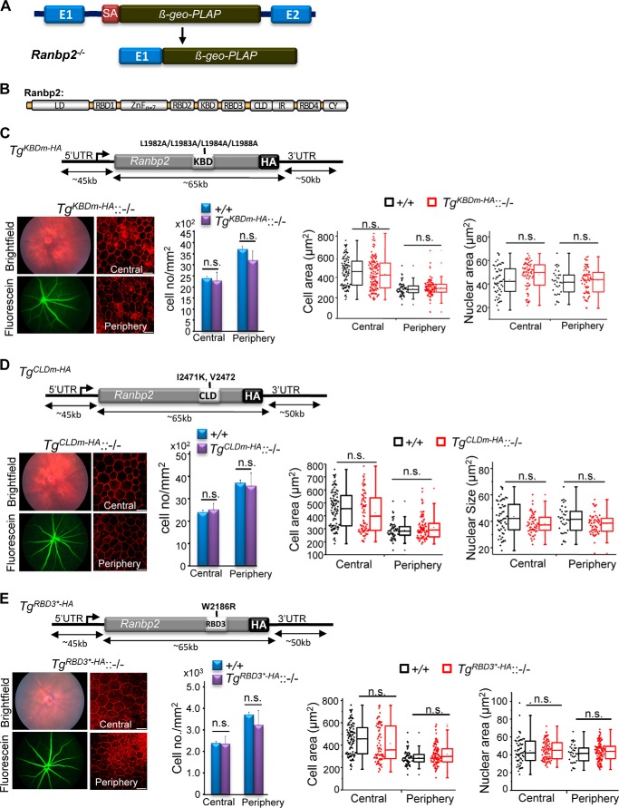
FIGURE 5.
Transgenic BAC constructs of Ranbp2 with loss-of-function mutations in KBD (TgKBDm-HA), CLD (TgCLDm-HA), or RBD3 (TgRBD3*-HA) expressed in mice with a Ranbp2−/− background (−/−) rescue RPE degeneration and mouse viability. A, constitutive disruption of Ranbp2 gene expression by the genomic insertion of a promoterless bicistronic (β-geo-PLAP) cassette with a splicing acceptor site (SA) between exons 1 and 2 leads to the splicing of exon 1 with the terminal bicistronic cassette. B, modular structure (domains) of Ranbp2. Selective mutations were introduced in various domains of Ranbp2 as shown in C–E. TgKBDm-HA::−/− (C), TgCLDm-HA::−/− (D), and TgRBD3*-HA::−/− mice (E) with expression of TgKBDm-HA, TgCLDm-HA, and TgRBD3*-HA, respectively, in a Ranbp2−/− (−/−) background have ordinary fundus and FITC-dextran angiograms, cell densities, and cellular and nuclear areas in the central and peripheral regions of the RPE at P35. The TgKBDm-HA, TgCLDm-HA, and TgRBD3*-HA constructs of Ranbp2 harbor the mutations L1982A/L1983A/L1984A/L1988A, I2471K/V2472, and W2186R, respectively. TgKBDm-HA::−/−, TgKBDm-HA::Ranbp2−/−; TgCLDm-HA::−/−, TgCLDm-HA::Ranbp2−/−; TgRBD3*-HA::−/−, TgRBD3*-HA::Ranbp2−/−; +/+, Ranbp2+/+. Data represent the mean ± S.D. (error bars) (cell density); dot-box plot analyses are shown for cellular and nuclear areas. Statistical analysis was done using a Mann-Whitney U test at a significance level of 0.05; n = 3, TgKBDm-HA::−/− and TgCLDm-HA::−/−; n = 4, TgRBD3*-HA::−/−; n.s., non-significant; LD, leucine-rich domain; RBDn = 1–4, Ran GTPase-binding domains, n = 1–4; ZnFn = 7, zinc finger-rich domains; IR1 + 2, internal repeat. Scale bars, 20 μm (C–E).

