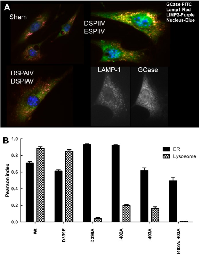
FIGURE 10.

Intracellular localization of GCase DSPIIV variants in Gba1−/− cells. A, typical examples of immunofluorescence localization of DSPIIV-substituted GCase variants following transient transfections. The DSPIIV (WT) and ESPIIV show co-localization of GCase (FITC) with either Lamp1 (red) or LIMP-2 (purple) (top right), indicating that the retention of charge by Glu399 does not impact localization. The cells are typical for either Asp399 (DSPIIV) or Glu399 (ESPIIV). The DSPAIV (I402A) or DSPIAV (I403A) mutant shows no co-localization with ldLIMP-2 or Lamp1 (bottom left) (i.e. no binding to ldLIMP-2 in the cell and no localization to the lysosome (Lamp1/ldLIMP2)). The black and white images in the bottom right indicate the relative localization of LAMP-1 (lysosomes) and WT GCase (ER/Golgi and lysosomes) in Gba1−/− fibroblasts for comparison. Sham control is shown in the top left. B, Pearson indices for co-localization of the various mutants in the ER/Golgi (black) or lysosome (hatched). The substitution of Glu for Asp (WT) at 399 (ESPIIV) did not alter the co-localization compared with WT. In comparison, ASPIIV (D399A) and the double mutant, DSPAAV (I402A/I403A) showed little co-localization to the lysosome but significant retention in the ER/Golgi. The single mutants, DSPAIV (I402A) and DSPIAV (I403A), showed partial co-localization to the lysosome but greater retention in the ER/Golgi. Error bars, S.E.
