FIGURE 3.

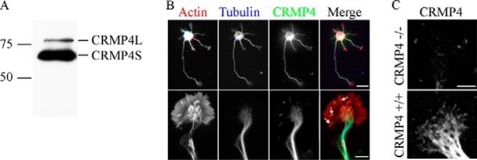
CRMP4 expression in hippocampal neurons. A, Western blot of an E18 wild-type hippocampal lysate probed with anti-CRMP4 antibody. B, 3 DIV wild-type hippocampal neurons stained with rhodamine phalloidin and anti-βIII-tubulin and anti-CRMP4 antibodies. CRMP4 is expressed in the microtubule-rich neurite and central domain and is also present in the actin-rich periphery (arrows). Scale bar, 20 (top row) and 5 μm (bottom row). C, 3 DIV hippocampal neurons from CRMP4+/+ or CRMP4−/− mice stained with anti-CRMP4 antibody. Scale bar, 5 μm.
