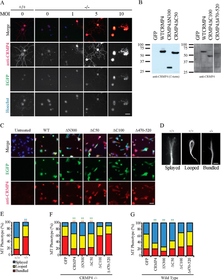
FIGURE 5.
CRMP4 promotes microtubule splaying through its tubulin-binding domain. A, hippocampal neurons from littermate control mice (+/+) or from CRMP4−/− mice transduced with an increasing multiplicity of infection (MOI) of wild-type CRMP4 virus were stained with anti-CRMP4 antibody. Neurons transduced with a multiplicity of infection of 5 generated wild-type levels of CRMP4 in neurites. Scale bar, 40 μm. B, Western blot of NIH 3T3 cells transduced with individual CRMP4 viruses at a multiplicity of infection of 5 and stained with an antibody raised against the carboxyl terminus (C-term) of CRMP4 (left panel) or a polyclonal CRMP4 antibody raised against full-length CRMP4 (right panel). Individual constructs were expressed at comparable levels. C, immunofluorescent staining of 3T3 cells transduced with individual CRMP4 constructs with a polyclonal CRMP4 antibody raised against full-length CRMP4. Scale bar, 50 μm. D, hippocampal neurons from CRMP4+/+ or CRMP4−/− mice were fixed, stained with anti-βIII-tubulin, and classified as having a splayed, looped, or bundled microtubule phenotype. Scale bar, 5 μm. E–G, quantitation of microtubule (MT) phenotype in hippocampal neurons from CRMP4+/+ and CRMP4−/− mice (E) or in CRMP4−/− growth cones transduced with HSV-GFP as a control or with HSV-CRMP4 rescue constructs (F) or for wild-type growth cones transduced with HSV-GFP as a control or with HSV-CRMP4 constructs (G). Determinations are mean ± S.E. from three experiments performed on 30 cells per experiment. **, p < 0.01 compared with GFP control condition by one-way ANOVA with Tukey's post hoc test. EGFP, enhanced GFP.

