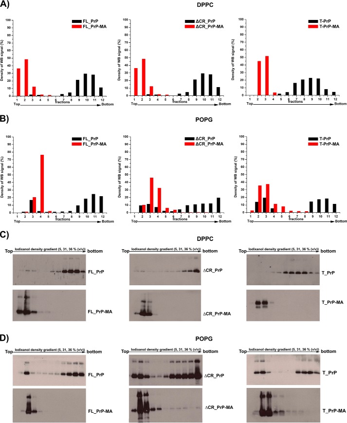
FIGURE 4.
Strong and specific binding of PrPs to liposomes via their C-terminal membrane anchors. The interactions of anchorless PrPs (black) and PrPs with a membrane anchor (red) with DPPC liposomes (A) and POPG liposomes (B) were analyzed by flotation assays in combination with Western blots. C and D, The original Western blots are depicted in C for experiments with DPPC liposomes and in D for POPG liposomes. POPG liposomes loaded with PrP variants were treated with a solution of 10 mm NaOH to disrupt electrostatic interactions. Quantification of all blots was performed by using ImageJ software.

