
Fig. 1.
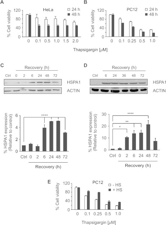
Heat shock preconditioning reduces thapsigargin-induced cytotoxicity in HeLa and PC12 cells. (A) HeLa cells and (B) PC12 cells were treated with indicated concentrations of TG for 24 h and 48 h followed by MTT assessment of cell viability. (C) HeLa cells and (D) PC12 cells were heat shocked in HEPES buffered media for 1 h at 42 °C and after recovery at 37 °C for indicated periods of time the induction of HSPA1 was determined by Western blotting (upper panel) using specific antibody against HSPA1. ACTIN was used as a loading control. Densitometric analysis (lower panel) of HSPA1 induction was normalized to ACTIN and expressed relative to untreated cells. Values shown are the mean ± SEM of three independent determinations. ∗P < 0.05, ∗∗P < 0.01, ∗∗∗∗P < 0.001 (E) PC12 cells were heat shocked or left untreated, and after a 6 h recovery period they were treated with increasing doses of TG for 48 h and an MTT assay was performed after that time. Average and error bars represent mean ± SD from two independent experiments.