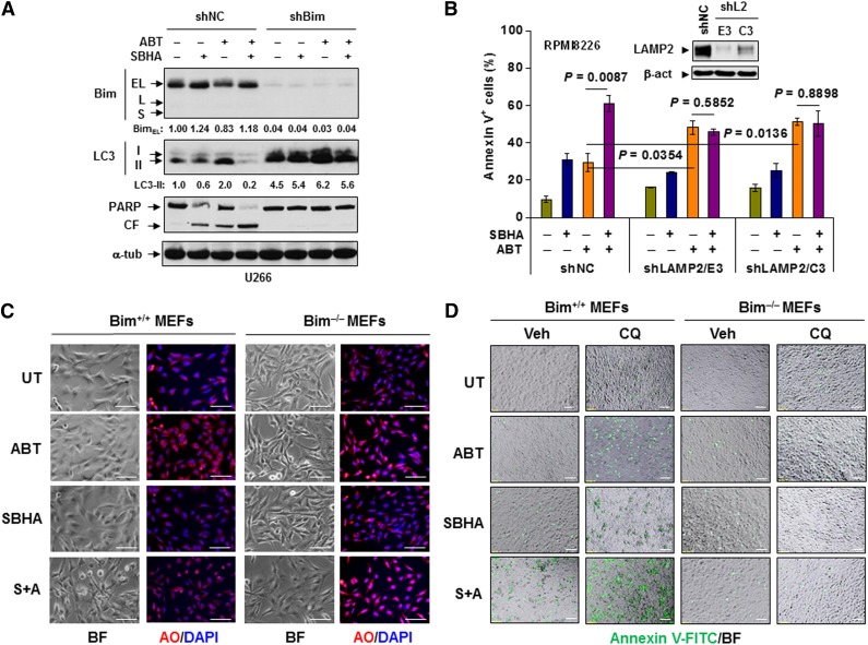
Figure 5.
Bim is required for disruption of autophagy and induction of apoptosis induced by ABT-737. (A) U266 cells stably transfected with Bim or scrambled sequence shRNA were incubated with 300 nM ABT-737 with or without 20 μM SBHA for 24 hours. Following treatment, immunoblotting analysis was performed to monitor Bim expression, LC3 processing, and PARP cleavage. (B) RPMI8226 cells stably transfected with LAMP2 (two subclones designated E3 and C3) or scrambled sequence shRNA (inset) were exposed to 300 nM ABT-737 with or without 20 μM SBHA for 24 hours followed by flow cytometry to determine the percentage of apoptotic cells. (C) MEFs derived from wild-type (Bim+/+) or bim knockout (Bim−/−) mice were treated with 500 nM ABT-737 with or without 20 μM SBHA for 16 hours, after which cells were stained with acridine orange (AO). (D) MEFs were exposed to 500 nM ABT-737 with or without 20 μM SBHA in the presence or absence of 50 μM CQ for 24 hours, after which cells were stained with annexin V-FITC. For both 5C and 5D images were captured by inverted fluorescence microscopy. BF, bright field. Scale bar = 10 μm; magnification ×10. S+A, SBHA + ABT-737; shL2, shLAMP2.

