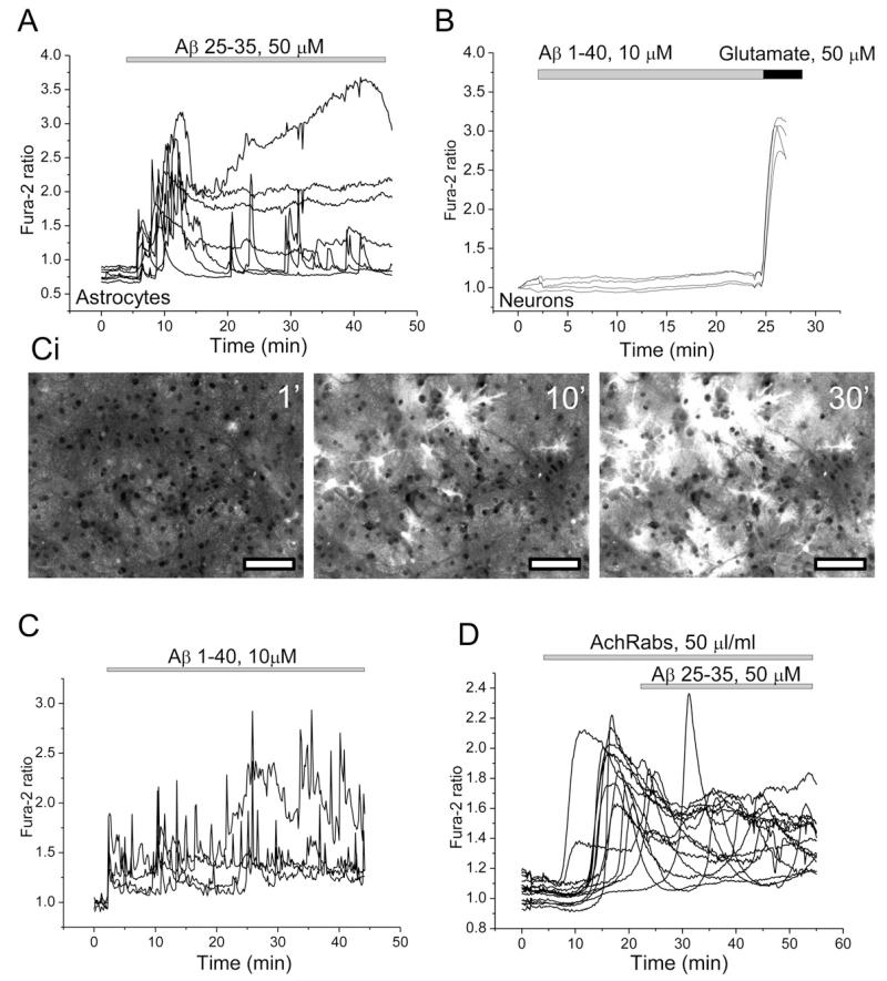
Fig. 1.
Aβ raises [Ca2+]c in astrocytes, effects of the antibodies on α7-type nAChRs. (A, C and D) Shows representative recordings of fura-2 ratio from astrocytes in hippocampal co-cultures following exposure to Aβ 25–35 (A) and Aβ 1–40 (C) peptides. Ci-representative images of fura-2 ratio under exposure to 10 μM Aβ 1–40. Bars on the images are 50 μm. The neurons showed no change in signal at all under application of amyloid (B). Their identity was confirmed by their response to glutamate (50 μM) at the end of the experiment. AchRabs, 50 μl/ml did not affect the calcium response to Aβ 25–35 in astrocytes (D). Each trace indicates [Ca2+]c measurement from a single cell.

