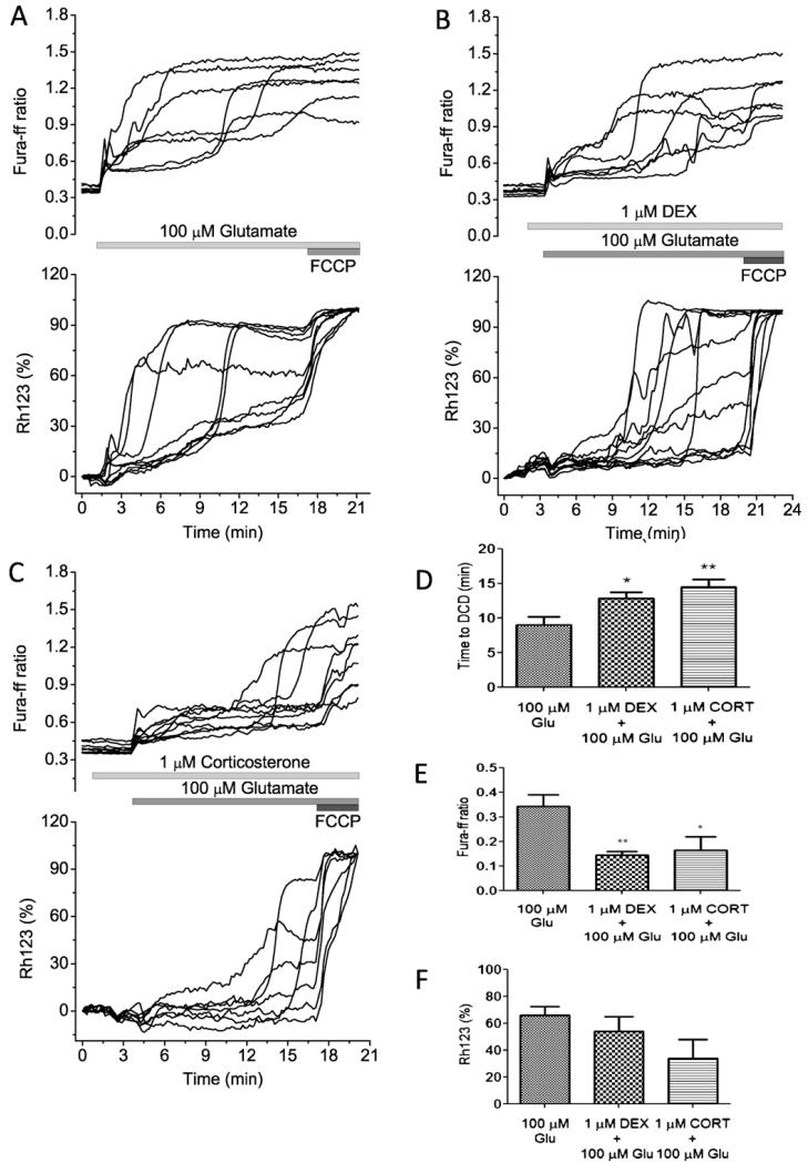
Fig. 6.
The effect of glucocorticoids on glutamate-induced calcium signal and mitochondrial membrane potential in co-cultured neurons and astrocytes. Simultaneous measurements of changes in [Ca2+]c (fura-ff ratio) and Δψm (relative Rh123 fluorescence) were made from single neurons. Glutamate (100 μM) and glycine (10 μM) were applied in a Mg2+-free solution. An increase in Rh123 fluorescence reflects mitochondrial depolarisation. (A) The calcium response to glutamate shows initial phase followed by calcium deregulation and loss of mitochondrial membrane potential. (B and C) The differences in the dynamics of glutamate-induced changes in [Ca2+]c and Δψm in the presence of 1 μM DEX or 1 μM corticosterone. The mitochondrial uncoupler FCCP (1 μM) was applied at the end of this and subsequent experiments to indicate the level of Rh123 fluorescence that reflected complete dissipation of Δψm. (D) Time (min) from application of glutamate and appearance of DCD; (E) differences in the amplitude (fura-ff ratio) of initial peak of glutamate (100 μM) induced calcium signal; (F) both glucocorticoids decrease the average of glutamate-induced Rh123 signal in neurons (*P < 0.05 and **P < 0.001).

