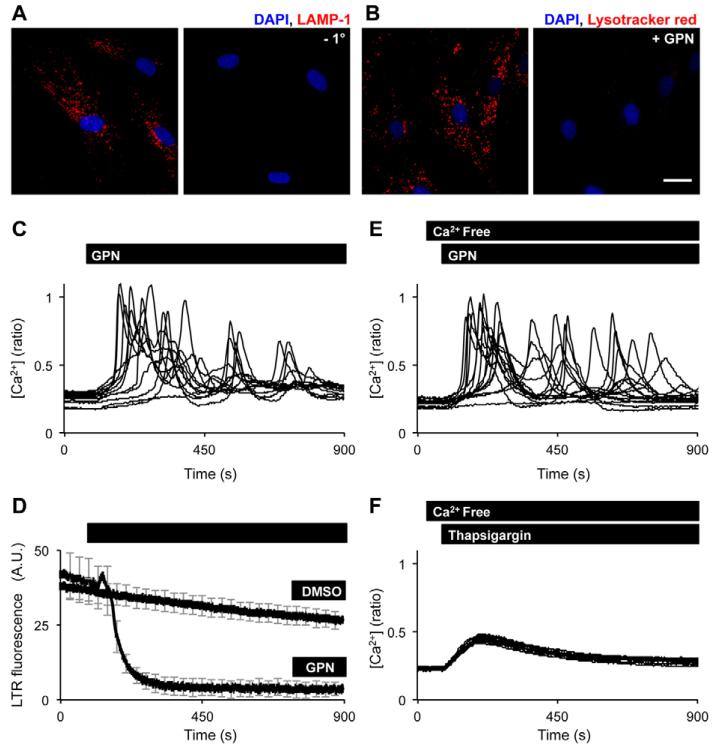
Fig. 1. Osmotic permeabilisation of lysosomes evokes complex Ca2+ signals.
(A) Confocal fluorescence image (red) of fibroblasts that were fixed and left unlabelled (right) or labelled (left) with a primary antibody raised to LAMP-1 and a far-red Alexa-Fluor-647-conjugated secondary antibody. (B) Confocal fluorescence images (red) of live fibroblasts that were labelled with Lysotracker Red before (left) or 216 seconds after (right) addition of 200 μM GPN. Nuclei were stained using DAPI (blue). Scale bar: 25 μm. (C,D) Single cell fluorescence responses recorded from cells loaded with either the Ca2+ indicator fura-2 (C) or Lysotracker Red (D) and stimulated with 200 μM GPN or the vehicle (DMSO). (E,F) Ca2+ signals in response to 200 μM GPN (E) or 1 μM thapsigargin (F) in the absence of extracellular Ca2+.

