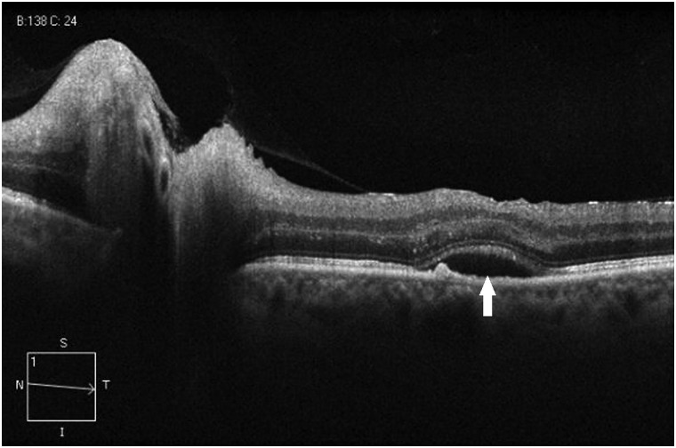
Figure 4.
Spectral domain optical coherence tomography line scan through the optic disc and macula showing subretinal fluid (arrow) in a middle-aged patient with a history of insulin-dependent diabetes mellitus, hypertension, hyperlipidaemia and sleep apnoea, who awoke with decreased vision in the left eye due to non-arteritic anterior ischaemic optic neuropathy.

