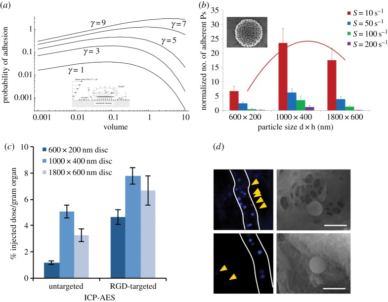
Figure 4.
Vascular adhesion of non-spherical nanoconstructs. (a) The probability of vascular adhesion grows as the shape deviates from spherical (γ = 1, sphere;  , quasi-discoidal particles) [2]. (b) Parallel plate flow chamber experiments showing a maximum vascular adhesion occurring for 1000 × 400 nm discoidal nanoconstructs [82]. (c) Tumour accumulation of untargeted and RGD-4C targeted discoidal nanoconstructs, demonstrating again a maximum accumulation for the 1000 × 400 nm discoidal nanoconstructs [44]. (d) Fluorescent images and scanning electron micrographs showing discoidal nanoconstructs (see yellow arrows) laying on the tumour neovasculature. 10% of the RBCs were stained in blue [44].

