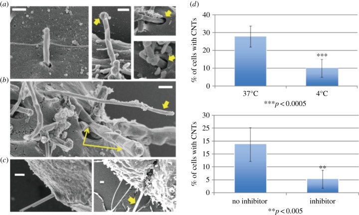
Figure 5.
Experimental observations of energy-dependent tip entry of one-dimensional nanomaterials into cells. (a) Carbon nanotubes entering murine liver cells. Arrow in middle panel shows carbon shell at the tube tip that distinguishes the nanotubes from surface microvilli. Arrows in right panels show close views of membrane invaginations surrounding the tubes at the point of entry. (b) Examples of nanotube tip entry in human mesothelial cells. Both an isolated tube (single arrow) and a tube bundle (double arrow) are seen in the process of high-angle entry. (c) Examples of active tip entry for other one-dimensional materials: 30 nm gold nanowires (left) and a 500 nm crocidolite asbestos fibre (right). (d) Effects of temperature and metabolic inhibitors on multi-walled CNT uptake as tests for active endocytic uptake. All images are obtained by field emission scanning electron microscopy following fixation and contrast enhancement with osmium tetroxide. All scale bars are 300 nm. Figure from [35].

