
Figure 2.
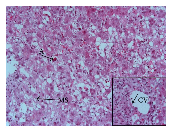
Histological view (haematoxylin-eosin stain, original magnification ×200) demonstrating extensive areas of hepatocytes with necrosis, ballooning degeneration, macro- and microvesicular steatosis (MS), and acidophil bodies (A). Insert: centrilobular vein (CV) showing endotheliitis (arrow) (original magnification ×400).