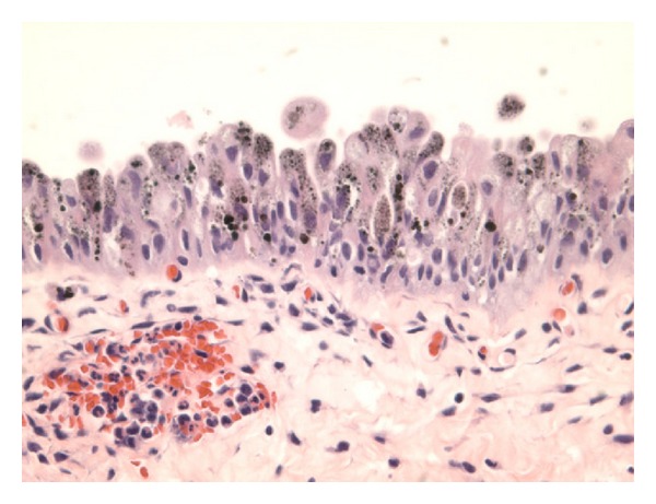
Figure 1.

Haematoxylin and Eosin staining showing melanosis and not melanoma: this shows light brown powdery pigments to dark brown globules which are mainly present in the cytoplasm, partially or completely obscuring the nuclei. There is no evidence of any pigment deposit in the lamina propria. Taken from Jin et al. [52].
