
Figure 7.
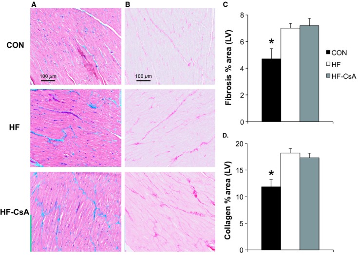
LV fibrosis and collagen deposition (N = 5 for each group). (A–B) representative histological sections of trichrome and Picrosirius red‐stained LV, showing increased fibrosis in HF and HF‐CsA animals. Magnification: ×40. (C–D) CsA treatment did not prevent an increase in total fibrosis and collagen induced by heart failure as indicated by assessment of percent area stained. (*P <0.05 vs. HF & HF‐CsA).