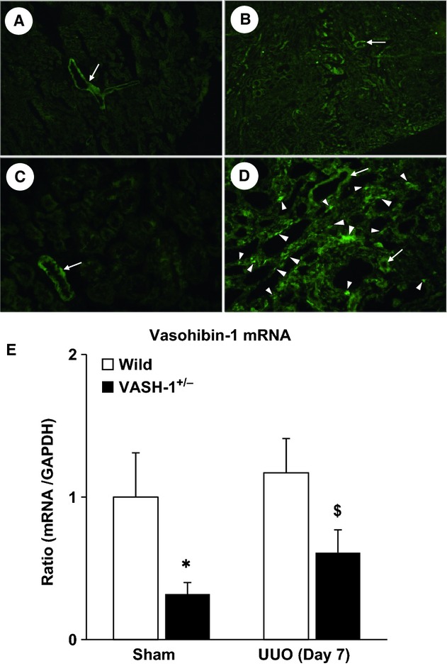
Figure 1.

Characterization of the VASH‐1 KO mice. (A–D) The VASH‐1 antibody was used to stain serial kidney sections from sham‐operated wild‐type (WT) (A, C) and WT/unilateral ureteral obstruction (UUO) (B, D) mice. (A, B) Original magnification; ×80. (C, D) Original magnification; ×400. (A, C) The immunoreactivity for VASH‐1 was mainly observed in the blood vessels (arrows). (B, D) The immunoreactivity for VASH‐1 was observed mainly in the renal interstitial cells (arrowheads), but was also observed in the blood vessels (arrows). (E) Real‐time PCR of WT (VASH1+/+) and VASH1+/− kidneys. The levels of VASH‐1 mRNA were normalized to those of GAPDH. The VASH‐1 mRNA levels were decreased in the VASH1+/− sham‐operated mice compared with the WT sham‐operated control mice. Similarly, the levels of VASH‐1 mRNA were decreased in the OBK of the VASH‐1+/− UUO mice compared with the WT UUO mice. OBK, obstructed kidneys.
