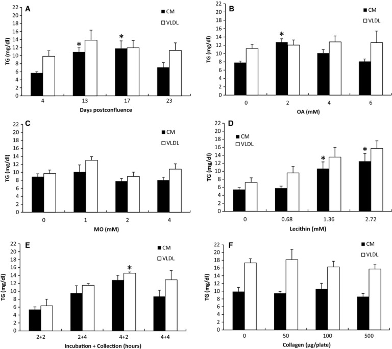
Figure 1.

The effect of cell differentiation, OA, MO, lecithin, incubation time, and collagen matrix on lipoprotein secretion. (A) Cell differentiation: cells that were 4, 13, 17, or 23 days postconfluent were incubated for 4 h with 4 mmol/L OA, 1 mmol/L MO, 0.68 mmol/L lecithin, and 1 mmol/L NaTC in growth media (n = 4 for each; except for 13 days, n = 3). (B) OA: Thirteen‐day postconfluent cells were incubated for 4 h with 0, 2, 4, or 6 mmol/L OA in growth media containing 1 mmol/L MO, 0.68 mmol/L lecithin, and 1 mmol/L NaTC (n = 4 for 0 and 4 mmol/L; n = 5 for 2 mmol/L; n = 3 for 6 mmol/L). (C) MO: incubated with 0, 1, 2, or 4 mmol/L MO in growth media containing 2 mmol/L OA, 0.68 mmol/L lecithin, and 1 mmol/L NaTC (n = 5 for 0 and 4 mmol/L; n = 3 for 1 mmol/L; n = 7 for 2 mmol/L). (D) lecithin: incubated with 0, 0.68, 1.36, or 2.72 mmol/L lecithin in growth media containing 2 mmol/L OA and 1 mmol/L NaTC (n = 3 for 0 and 0.68 mmol/L; n = 4 for 1.36 and 2.72 mmol/L). (E) Time: incubated with 2 mmol/L OA, 1.36 mmol/L lecithin, and 1 mmol/L NaTC in growth media for either 2 or 4 h; and collected for either 2 or 4 h in growth media (n = 3 for 2 + 2 and 4 + 2; n = 4 for 2 + 4 and 4 + 4). (F) Collagen: Each 10 cm tissue culture dish was coated with 0, 50, 100 or 500 μg collagen before it was populated with cells (n = 3 for each; except 0 μg, n = 4). After the cells have reached 13‐day postconfluence, they were incubated for 4 h with 2 mmol/L OA, 1.36 mmol/L lecithin, and 1 mmol/L NaTC in growth media. The secreted lipoproteins were then collected for 2 h in growth media, and separated into CM and VLDL layers by sequential NaCl density gradient ultracentrifugation. The concentration of TG in the CM and VLDL layers was measured. Refer to Table 1 for the schematic representation of the experimental conditions. There were at least three separate experiments performed for each study. n represents the total number of dishes. All of the TG measurements were done in duplicates. Mean ± SE. One‐way ANOVA with multiple comparison tests was used (*P <0.05 when compared with the groups without *. Comparisons were made only among the CM samples; among the VLDL samples; not between the CM and the VLDL samples).
