Figure 3.
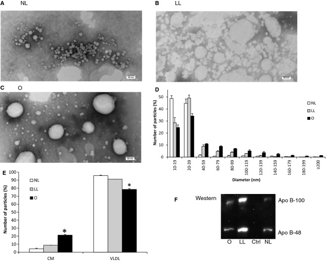
Comparing lipoprotein size and ApoB‐48 generated under the optimal and the previously reported conditions. Cells were grown on Transwell until they reached 13‐day postconfluence. The apical compartment was added with growth media (NL group); 1.6 mmol/L OA and 0.5 mmol/L NaTC in growth media (LL group); or 2 mmol/L OA, 1.36 mmol/L lecithin, and 1 mmol/L NaTC in growth media (O group); and the basolateral compartment was concurrently added with the growth media. After incubating the cells for either 4 (O and NL groups) or 14 h (LL group), the basolateral media was subjected to one‐step NaCl density gradient ultracentrifugation. The isolated lipoproteins from NL (A), LL (B), and O groups (C) were negatively stained, and their representative TEM images were depicted (scale bar = 80 nm). The size distribution of their lipoproteins was displayed in a histogram (D). The relative percentage of the number of CM particles (80 nm or larger) to the number of VLDL particles (smaller than 80 nm) was also depicted (E). Ten microliters of the lipoprotein fraction from each of the groups (from left: O, LL, growth media control, NL groups) was run on 4–20% polyacrylamide gradient gel under the reducing condition, transferred to nitrocellulose membrane, and blotted for ApoB. The top bands were ApoB‐100 and the bottom bands were ApoB‐48 (F). Two separate experiments were performed. Mean ± SE. One‐way ANOVA with multiple comparison tests was used (*P <0.05).

