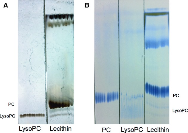
Figure 6.

Determining the presence of lysoPC in egg lecithin by TLC. (A) 0.5 mg of lysoPC (left) and 2 mg egg lecithin (right) in chloroform were separated on silica gel 60 plates using chloroform:methanol:acetic acid:water (50:37.5:3.5:2) (v:v:v:v) as the solvent system. The plate was stained by choline staining followed by 20% sulfuric acid staining with 100°C heating until the color developed. (B) 0.5 mg PC (left), 5 mg lysoPC (middle), 0.5 mg lecithin (right) in chloroform:methanol:water (12:7:1) (v:v:v) were separated on silica gel 60 plates using chloroform:ethanol:water:triethylamine (30:35:7:3.5) (v:v:v:v) as the solvent system. The plate was stained by PL staining with 100°C heating until the color developed.
