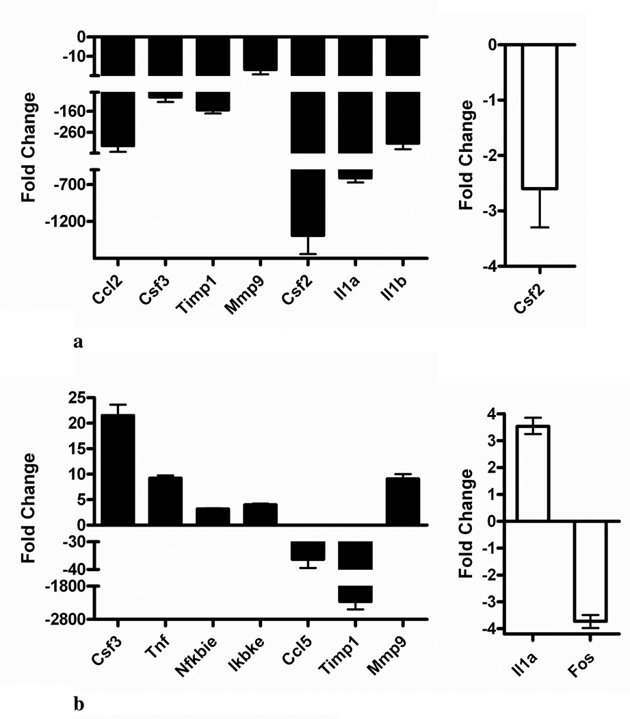
Figure 2.
Quantitative real-time RT-PCR validation of microarray results. RAW 264.7 cells were stimulated with 0.1 µg/mL LPS and untreated or treated (A) for 6 h or (B) for 24 h with either 35 µM HNE (black bars) or 0.1 mg/mL BH (white bars). Fold-changes (treated, stimulated cells relative to stimulated controls) are shown (X̄ ± 99% confidence interval for quadruplicate measurements of n = 3 biological replicates). Abbreviations: chemokine (C-C motif) ligand (Ccl); colony stimulating factor (Csf); tissue inhibitor of metalloproteinase 1 (Timp1); matrix metalloproteinase 9 (Mmp9); interleukin 1 (Il1); tumor necrosis factor (Tnf); nuclear factor of kappa light polypeptide gene enhancer in B-cells inhibitor, epsilon (Nfkbie); inhibitor of kappaB kinase, epsilon (Ikbke); and FBJ osteosarcoma oncogene (Fos).

