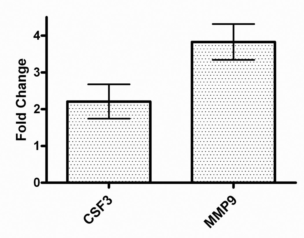
Figure 3.
ELISA validation of microarray results. Equivalent numbers of cells (4 × 10⁶/well) were stimulated with LPS (1 µg/mL) in the absence or presence of 35 µM HNE for 24 h. CSF3 (colony stimulating factor 3 (granulocyte)) and MMP-9 (matrix metalloproteinase 9) released into the culture medium were analyzed by ELISA. Fold changes (HNE-treated stimulated cells relative to stimulated controls) are shown (X̄ ± SD for triplicate measurements, representative of three independent experiments).

