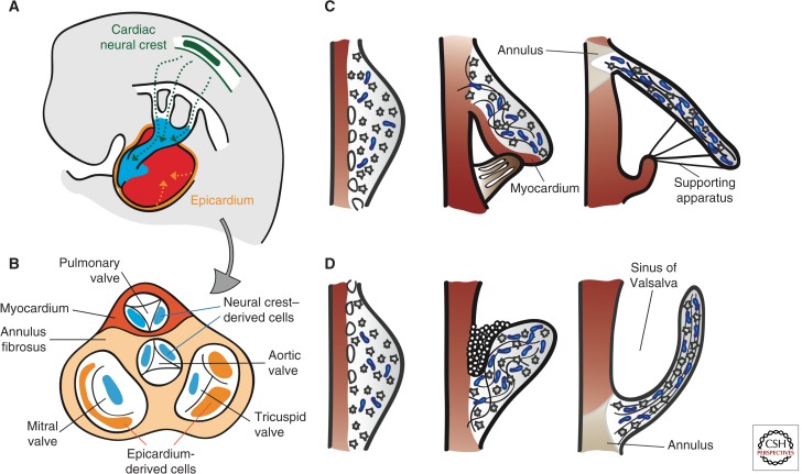
Figure 4.
Remodeling of the cardiac valve leaflets and cusps. (A) Contribution of cardiac neural crest (green)- and epicardial (orange)- derived cells (CNCC and EPDCs). CNCCs migrate through the pharyngeal arches and into the OFT to initiate the reorganization of the OFT and formation of SL valves. EPDCs contribute to the formation of the coronary arteries, interstitial cells in the myocardium, and the AV valves. (B) Top view of the septated heart, depicting the relative contributions of CNCCs and EPDCs to cardiac valve leaflets and cusps. (C) Morphogenesis of the AV valves. From E13.5 onward, the leaflets and tensile apparatus (light brown) of the AV valves form predominantly by delamination of the inner layers of the inlet zone from the ventricular septal wall. The chordae tendinae, annulus fibrosus, and the leaflet itself are derived from the endocardium. The septal leaflet of the TV (in contrast to the aortic leaflet of the MV), together with its supporting tendinous cords, remains connected to the myocardium until E17.5. (D) Morphogenesis of the SL valves. The excavation of the cusps takes place initially by solid ingrowth of the endothelium at the arterial face of the cusp and, subsequently, by lumenation. Elongation and remodeling of the primordia into mature valve structures are associated with regionalized cell proliferation and matrix alignment guided by hemodynamic forces. Myocardium is depicted in dark red, proteglycans in blue, and mesenchyme in gray.

