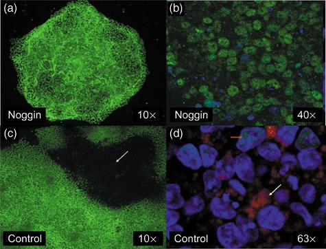
Figure 5.

Effect of noggin on colony morphology and protein expression. Cells were cultured in irradiated mouse embryonic fibroblast conditioned medium on Matrigel‐coated chamber slides in the absence (c and d) or presence of noggin (a and b). Human embryonic stem cell colonies cultured in the presence of noggin for 7 days and stained with (a) an Oct4 antibody [fluorescein isothiocyanate (FITC)] or (b) stained for Oct4 (FITC) and hCGβ (phycoerythrin) and counterstained with DAPI. (c) Spontaneously differentiating colony grown on Matrigel showing typical doughnut‐shaped appearance with Oct4‐negative, differentiated cells in the centre of the photograph (dark area indicated by arrow). (d) Spontaneously differentiated cells stained for Oct4 (FITC), and hCGβ (phycoerythrin) and counterstained with DAPI. Orange arrow indicating residual nuclear Oct4 staining (green) in some cells and also much larger nuclei (DAPI) of the differentiated cells. White arrow is shows positive hCGβ (red) staining.
